Substernal goiter (or retrosternal goiter) is an enlarged thyroid gland with intrathoracic extension. It remains unclear which goiters are to be termed substernal, but a recently proposed definition is a goiter that requires mediastinal exploration and dissection for complete removal or an intrathoracic component extending >3 cm in the thoracic inlet
Chest x-ray may show a superior mediastinal radiopacity causing the deviation of trachea to the opposite side. The superior margin of the radiopacity/mass is untraceable (cervicothoracic sign).
On ultrasound, the inability to scan the inferior most of the thyroid due to its extension posterior to the sternum makes substernal thyroid likely.
The most important CT features in determining the necessity of sternotomy for goiter excision are the presence of an ectopic goiter, total thyroid gland volume and goiter extension below the tracheal carina Most anterior substernal thyroid goiters are accessed via a transcervical approach. For goiters that cannot be removed via neck dissection, such as those with complicated anatomic extensions or posterior mediastinal involvement, the surgeon may need to incorporate a partial upper sternotomy and clavicular head resection or mini thoracotomy for adequate exposure.
thyroxine. 99mTc-thyroid images were heterogeneous, sub-optimal and difficult to interpret, especially if the patient on
In such situation, 99mTc Sestamibi thyroid planar & hybrid SPECT-CT imaging is useful because of different mechanism of the tracer uptakes and better quantity of images.
Hybrid SPECT-CT images show exact topography and size of mass and the type of underlying morphological changes that have occurred within the thyroid gland. the diagnostic value of SPECT-CT in retrosternal goiter (as demonstrated in this case).
63 years old lady with history of pressure symptoms at lower neck and sob on exertion. diagnosed as cardiac case but later on excluded.
Past history of large multinodular goiter underwent near total thyroidectomy with retrosternal extension couple of years ago and on 50 microgram thyroxine per day.
FT3 1.35 nmol/L (58.1-140.2), FT4 108.4 nmol/L, TSH 0.69 micro-IU/ml (0.27- 4.2
Evidence of lobulated soft tissue density, mildly enhancing lesion approximately 0f 4.5*3.6 cm noted at the cervicothoracic junction anteriorly (pre tracheal) region extending from the neck into the superior mediastinum with tiny foci of calcification within it, possibly lymph node mass
(differential diagnosis can be ? exophytic thyroid nodule extending into superior mediastinum). It is mildly displacing the trachea of right side. Posterior to this lesion evidence of coarse nodular calcification approximately of 9.5*6.3 mm in size noted (possibly calcified lymph node). Multiple enlarged mediastinal and hilar lymph nodes noted. Calcific densities in both the hilar regions.
The right lobe is seen with decreased and patchy tracer uptakes. Large area of decreased and patchy tracer uptake seen inferior to the left thyroid bed with retrosternal extension (Better appreciated in HYBRID SPECT CT IMAGES).
Also, linear shaped abnormal tracer uptake seen in lower esophageal region (Better appreciated in HYBRID SPECT CT IMAGES (Most likely to be physiological tracer hold).
It shows visualization of right lobe with decreased tracer uptakes. Large area (lobulated, which has bigger and smaller component) of abnormal MIBI tracer uptake seen inferior to the left thyroid bed, which is partially extending retrosternal (better appreciated in HYBRID SPECT CT IMAGES).
As underlying CT images shows bi-lobed lesion arising from the left lobe of the thyroid gland. a) The bigger component is predominantly solid with punctate calcfic foci. b) The smaller component shows an inferior extension retrosternal (approximately 1.5 cm below the plane of thoracic inlet (Top of the manubrium sternum to the superior border of the D1 vertebra) and the lesion is above the arch of aorta. c) The approximate measurement of the mass is 5.6 cms antero-posterior and transverse 3.5 cm in diameter)
THE HYBRID SPECT CT SHOWS TRACER UPTAKES IN LEFT LOBE MASS WITH RETROSTERNAL EXTENSION ON BOTH 99mTc Pertechnetate & 99mTc Sestamibi thyroid images) AND HENCE IT IS LIKELY TO BE LEFT LOBE THYROIDAL MASS WITH PARTIAL RETROSTERNAL EXTENSION (1.5 cm below the plane of thoracic inlet and it is above the arch of aorta).
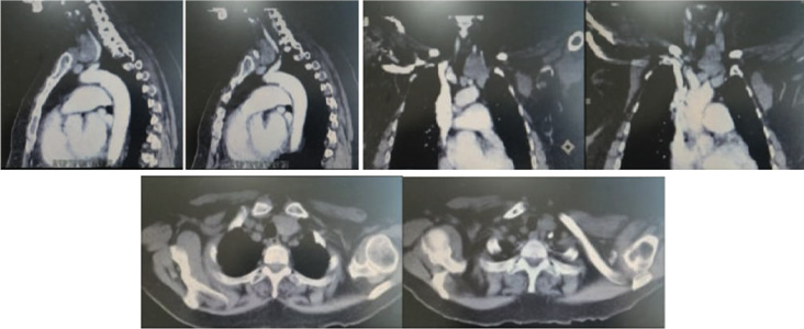
CT scan images
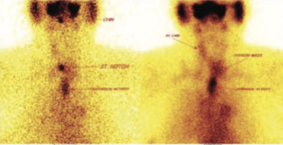
99mTc pertechnatate thyroid images
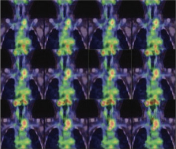
99mTc pertechnatate hybrid SPECT CT images showing left lobe mass with retrosteranl extension
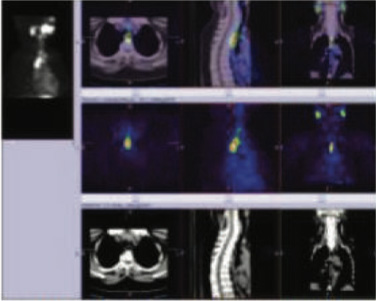
9mTc pertechnatate hybrid SPECT CT images showing esophageal activity
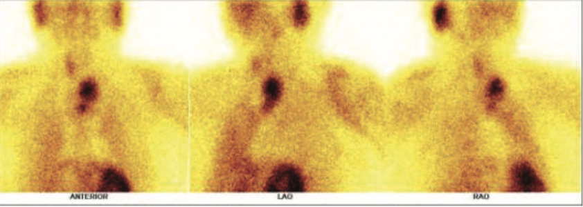
99mTc SESTAMIBI thyroid images showing left lobe thyroid mass with retrosternal uptakes

99mTc Sestamibi hybrid SPECT CT images showing left lobe thyroid mass with retrosteranl extension
Scintigraphy diagnosis of retrosternal goiter plays an important role in avoiding invasive diagnostic procedures, and hybrid SPECT-CT acquisition may enhance the diagnostic sensitivity of this technique.
Hybrid SPECT-CT provided additional diagnostic information for the successful treatment of patient with retrosternal goiter and thyroid nodularity.
A surgeon with an understanding of the radiologic reporting of a substernal goiter on a dedicated chest CT might perform a sternotomy instead of a simple low-collar incision for resection of substernal goiter.
Some suggested imaging features may indicate requirement for a thoracic approach with a sternotomy include 8